
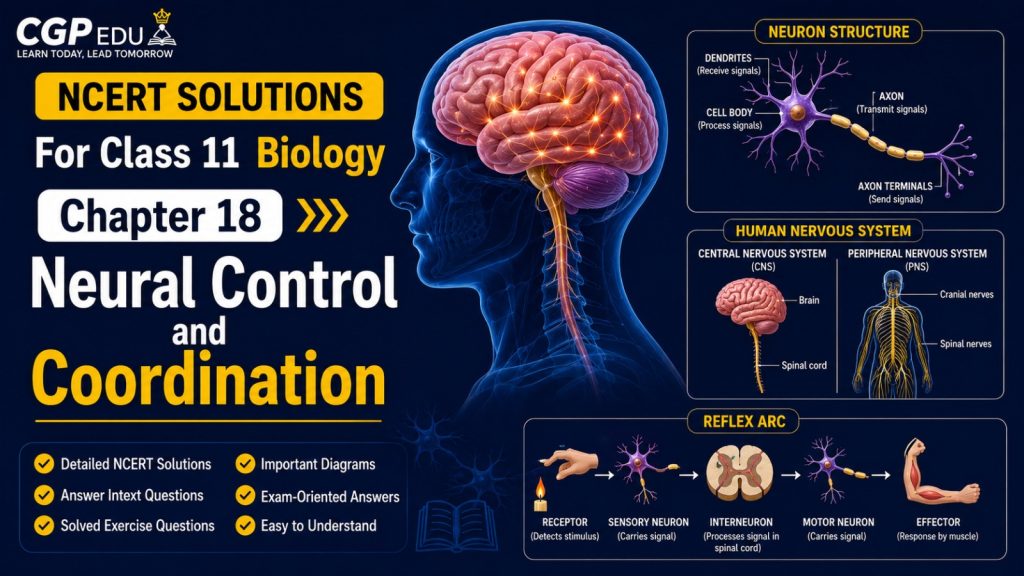
The chapter Neural Control and Coordination introduces students to the neural system of animals and explains how neural control and coordination are achieved in the human body. It covers the central nervous system, peripheral nervous system, neuron structure, generation and conduction of nerve impulses, synaptic transmission, and sensory organs like the eye and ear.
Students learn about the structure and types of neurons, resting potential, action potential, depolarisation, repolarisation, and the propagation of nerve impulses. The chapter also explains the structural organisation of the human brain, spinal cord, reflex action, reflex arc, and the working of specialized sensory receptors for vision and hearing. A clear understanding of Neural Control and Coordination provides a strong foundation for studying human physiology, nervous coordination, sensory perception, and integrative functions, making it an important chapter for NEET preparation.
Class 11 Biology Chapter 18 Overview
Students learn about the human neural system, including the central neural system ($\text{CNS}$) and peripheral neural system ($\text{PNS}$), along with the structure and types of neurons. The chapter covers the generation and conduction of nerve impulses, synaptic transmission across chemical synapses, and the anatomical organisation of the brain and spinal cord.
Students are introduced to the forebrain, midbrain, hindbrain, cerebrum, thalamus, hypothalamus, cerebellum, pons, and medulla oblongata. The chapter also covers reflex action, reflex arcs, and detailed structures of sensory organs such as the eye (cornea, lens, retina, rods, and cones) and the ear (external, middle, and inner ear, cochlea, and organ of Corti). A clear understanding of Neural Control and Coordination helps students develop a strong foundation in human physiology, neural regulation, sensory processing, and homeostasis, making it an important chapter for NEET preparation.
NCERT Solutions for Class 11 Biology
Chapter 18 – Neural Control And Coordination
Question 18.1. Briefly describe the structure of the following:
(a) Brain (b) Eye (c) Ear
Solution:
(a) Brain: The brain acts as control and command system of the body. It is protected by skull and is covered by three meninges. It is divisible into three main regions: forebrain, midbrain and hindbrain.
(i) Forebrain – It consists of three regions:
– Olfactory lobes: These are a pair of very small, solid club-shaped bodies which are widely separated from each other. They are fully covered by cerebral hemispheres.
– Cerebrum – It is the largest and most complex of all the parts of human brain. A deep cleft divides the cerebrum into right and left cerebral hemispheres, connected by myelinated fibres, the corpus callosum.
– Diencephalon – It encloses a slit-like cavity, the third ventricle. The thin roof of this cavity is known as the epithalamus, the thick right and left sides as the thalami, and floor as the hypothalamus.
(ii) Midbrain – It is located between thalamus/hypothalamus of forebrain and pons of hindbrain. Its upper surface has two pairs of rounded protrusions called corpora quadrigemina and two bundles of fibres called crura cerebri.
(iii) Hindbrain – It consists of:
– Cerebellum – The second largest part of the human brain is the cerebellum. It consists of two lateral cerebellar hemispheres and central worm-shaped part, the vermis.
– Pons varolii – An oval mass lying above the medulla oblongata, consisting mainly of nerve fibres which interconnect different regions of the brain.
– Medulla oblongata – It extends from the pons varolii above and is continuous with the spinal cord below. The midbrain, pons varolii and medulla oblongata are collectively called brain stem.
(b) Eye: Eye is a hollow spherical structure composed of three coats:
(i) Fibrous coat: It has two distinct regions – sclera and cornea. Sclera covers most of the eyeball and contains collagen fibres. Cornea is a transparent portion that forms the anterior one-sixth of the eyeball.
(ii) Vascular coat: Comprises choroid, iris, and ciliary body.
(iii) Nervous coat: Consists of retina which is the neural and sensory layer containing ganglion cells, bipolar cells and photoreceptor cells (rods and cones).
(c) Ear: There are three portions in an ear:
(i) External ear: Consists of pinna and external auditory canal or meatus.
(ii) Middle ear: Consists of 3 small bones called ear ossicles – malleus, incus and stapes.
(iii) Internal ear: Consists of bony and membranous labyrinth, including three semicircular ducts, utricle, saccule and cochlea.
Question 18.2. Compare the following:
(a) Central neural system (CNS) and Peripheral neural system (PNS).
(b) Resting potential and action potential.
(c) Choroid and retina.
Solution:
(a) CNS and PNS:
– CNS lies along the mid-dorsal axis of the body, comprising the brain and spinal cord, serving as the center of information processing and control.
– PNS consists of nerves arising from the CNS (cranial and spinal nerves) that carry information to and from the CNS.
(b) Resting potential and action potential:
– Resting potential is the potential difference across the plasma membrane of a resting nerve fibre, where the outside is positively charged relative to the inside (electrically polarized).
– Action potential is the reversal of polarity across the plasma membrane (depolarization) during the excited state when the inside becomes positively charged relative to the outside.
(c) Choroid and retina:
– Choroid lies adjacent to the sclera, contains numerous blood vessels to supply nutrients and oxygen, and is pigmented dark brown.
– Retina is the inner neural and sensory layer of the eyeball containing photoreceptor cells (rods and cones), bipolar cells, and ganglion cells.
Question 18.3. Explain the following processes:
(a) Polarisation of the membrane of a nerve fibre.
(b) Depolarisation of the membrane of a nerve fibre.
(c) Conduction of a nerve impulse along a nerve fibre.
(d) Transmission of a nerve impulse across a chemical synapse.
Solution:
(a) Polarisation: In resting nerve fibres, $\text{Na}^+$ ions predominate outside while $\text{K}^+$ ions predominate inside. The differential ion permeability and active transport create a positive charge outside and a negative charge inside the membrane, making it polarized.
(b) Depolarisation: When a stimulus is applied, $\text{Na}^+$ channels open, causing $\text{Na}^+$ ions to rush into the axon. This reverses the polarity (inside becomes positive relative to outside), resulting in depolarisation.
(c) Conduction of a nerve impulse: An action potential generated at one spot on the nerve fibre is self-generated and propagated as a wave along the entire length of the axon.
(d) Transmission across a chemical synapse: When an action potential reaches the axon terminal, synaptic vesicles fuse with the presynaptic membrane and release neurotransmitters into the synaptic cleft. These neurotransmitters bind to specific receptors on the postsynaptic membrane, opening ion channels and generating a new potential in the postsynaptic neuron.
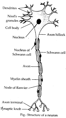
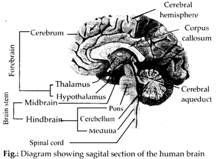
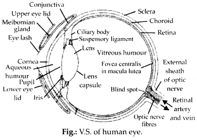
Question 18.4. Draw labelled diagrams of the following:
(a) Neuron (b) Brain
(c) Eye (d) Ear
Solution:
a.
b.
c.
d.
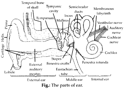
Question 18.5. Write short notes on the following:
(a) Neural coordination (b) Forebrain
(c) Midbrain (d) Hindbrain
(e) Retina (f) Ear ossicles
(g) Cochlea (h) Organ of Corti
(i) Synapse
Solution:
(a) Neural coordination: The working together of various organs of a multicellular organism in a systematic manner to complement each other’s functions, achieved through sensory input, integration, and motor output.
(b) Forebrain: Consists of olfactory lobes, cerebrum (divided into hemispheres connected by corpus callosum), and hypothalamus/thalamus.
(c) Midbrain: Located between forebrain and hindbrain, contains corpora quadrigemina on its dorsal portion.
(d) Hindbrain: Comprises pons, cerebellum, and medulla oblongata, controlling vital reflexes and balance.
(e) Retina: Inner layer of the eye containing photoreceptor cells (rods for twilight vision, cones for daylight and colour vision).
(f) Ear ossicles: Chain of three bones in the middle ear – malleus, incus, and stapes – that amplify sound wave transmission.
(g) Cochlea: Spirally coiled structure of the inner ear responsible for hearing.
(h) Organ of Corti: Located on the basilar membrane inside the cochlea, containing hair cells that act as auditory receptors.
(i) Synapse: Junction between the axon of one neuron and the dendrite/cyton of another neuron for impulse transmission.
Question 18.6. Give a brief account of
(a) Mechanism of synaptic transmission.
(b) Mechanism of vision.
(c) Mechanism of hearing.
Solution:
(a) Refer to Solution 18.3(d).
(b) Mechanism of vision: Light rays focus on the retina, inducing dissociation of retinal from opsin, which alters membrane permeability and generates receptor potentials. This triggers action potentials in ganglion cells via bipolar cells, which are transmitted via optic nerves to the visual cortex.
(c) Mechanism of hearing: Sound waves strike the tympanum, producing vibrations transmitted via ear ossicles to the oval window, creating fluid waves in the cochlea. These waves stimulate hair cells in the organ of Corti against the tectorial membrane, generating nerve impulses transmitted via auditory nerves to the brain.
Question 18.7. Answer briefly.
(a) How do you perceive the colour of an object?
(b) Which part of our body helps us in maintaining the body balance?
(c) How does the eye regulate the amount of light that falls on the retina?
Solution:
(a) Through three types of cone cells in the retina containing photopigments sensitive to red, green, and blue light. Equal stimulation produces white light sensation, while combinations produce various colours.
(b) Ears (specifically cristae and maculae in the vestibular apparatus of the inner ear).
(c) By the iris, which contains circular sphincter muscles (making the pupil smaller in bright light) and radial dilator muscles (widening the pupil in dim light).
Question 18.8. Explain the following.
(a) Role of $\text{Na}^+$ in the generation of action potential.
(b) Mechanism of generation of light-induced impulse in the retina.
(c) Mechanism through which a sound produces a nerve impulse in the inner ear.
Solution:
(a) $\text{Na}^+$ ions rush into the axon upon stimulus, decreasing membrane potential, reversing polarity (depolarization), and peaking at $+40$ to $+50\text{ mV}$ to form the action potential.
(b) Refer to Solution 18.6(b).
(c) Refer to Solution 18.6(c).
Question 18.9. Differentiate between
(a) Myelinated and non-myelinated axons
(b) Dendrites and axons
(c) Rods and cones
(d) Thalamus and Hypothalamus
(e) Cerebrum and Cerebellum
Solution:
(a) Myelinated vs Non-myelinated axons:
| Myelinated axon | Non-myelinated axon |
|---|---|
| Enveloped with myelin sheath with nodes of Ranvier. | Not enclosed by myelin sheath; continuous conduction. |
| Faster impulse conduction (saltatory). | Slower impulse conduction. |
(b) Dendrites vs Axons:
| Dendrites | Axons |
|---|---|
| Short, tapered fibres carrying impulses towards the cell body. | Long fiber carrying impulses away from the cell body. |
(c) Rods vs Cones:
| Rods | Cones |
|---|---|
| Responsible for dim light (scotopic) vision; contain rhodopsin. | Responsible for bright light and colour (photopic) vision. |
(d) Thalamus vs Hypothalamus:
| Thalamus | Hypothalamus |
|---|---|
| Acts as a relay station for sensory and motor impulses. | Controls body temperature, urge for eating, drinking, and hormone secretion. |
(e) Cerebrum vs Cerebellum:
| Cerebrum | Cerebellum |
|---|---|
| Largest part of the brain; controls intelligence, memory, and voluntary actions. | Second largest part; coordinates posture, balance, and muscle synergy. |
Question 18.10. Answer the following.
(a) Which part of the ear determines the pitch of a sound?
(b) Which part of the human brain is the most developed?
(c) Which part of our central neural system acts as a master clock?
Solution:
(a) The receptor cells in the organ of Corti (internal ear).
(b) Cerebrum (cerebral hemispheres).
(c) Pineal gland (associated with diencephalon of forebrain) acts as a master clock maintaining biological rhythms.
Question 18.11. The region of the vertebrate eye, where the optic nerve passes out of the retina, is called the
(a) fovea (b) iris
(c) blind spot (d) optic chiasma
Solution:
(c) blind spot
Question 18.12. Distinguish between
(a) Afferent neurons and efferent neurons
(b) Impulse conduction in myelinated nerve fibre and unmyelinated nerve fibre
(c) Aqueous humour and vitreous humour
(d) Blind spot and yellow spot
(e) Cranial nerves and spinal nerves
Solution:
(a) Afferent vs Efferent neurons:
| Afferent neurons | Efferent neurons |
|---|---|
| Transmit impulses from sensory organs to CNS. | Transmit impulses from CNS to effector organs. |
(b) Refer to Solution 18.9(a).
(c) Aqueous vs Vitreous humour:
| Aqueous humour | Vitreous humour |
|---|---|
| Watery fluid present in the anterior chamber between cornea and lens. | Jelly-like fluid present in the posterior chamber between lens and retina. |
(d) Blind spot vs Yellow spot (Fovea):
| Blind spot | Yellow spot (Fovea) |
|---|---|
| Region where optic nerve leaves retina; devoid of photoreceptors. | Point of greatest visual acuity containing densely packed cones. |
(e) Cranial vs Spinal nerves:
| Cranial nerves | Spinal nerves |
|---|---|
| Nerves arising directly from the brain. | Nerves arising from the spinal cord. |
Why Class 11 Biology Chapter 18 Matters in NEET
Class 11 Biology Chapter 18: Neural Control and Coordination is highly important for NEET because it explains how the nervous system receives, processes, and responds to information from the internal and external environment. The chapter covers important concepts such as neurons, nerve impulses, the human nervous system, brain, spinal cord, reflex action, synaptic transmission, and sensory organs. NEET frequently includes NCERT-based questions on the structure and functions of neurons, nerve impulse transmission, synapses, the central and peripheral nervous systems, brain regions, reflex arcs, and the structure and functioning of the eye and ear. A thorough understanding of this chapter helps students build a strong foundation in human physiology, nervous coordination, sensory perception, and the integration of body functions.
Preparation Tips for Class 11 Biology Chapter 18
Begin by understanding the neural system and types of nervous systems found in different organisms. Learn the basic organisation of the human nervous system and understand the differences between the central nervous system, peripheral nervous system, and autonomic nervous system.
Study the structure of a neuron carefully, including the cell body, dendrites, axon, myelin sheath, nodes of Ranvier, and axon terminals. Understand the difference between sensory, motor, and interneurons and learn how neurons transmit information.
Pay special attention to the generation and conduction of nerve impulses. Understand the concepts of resting potential, action potential, depolarisation, repolarisation, and the role of sodium and potassium ions. Revise how an impulse travels along the axon and reaches the axon terminal.
Study synaptic transmission thoroughly. Understand the structure and function of chemical synapses, neurotransmitter release, synaptic cleft, and the transmission of signals from one neuron to another. Also revise the difference between electrical and chemical synapses.
Learn the organisation and functions of the human brain and spinal cord. Pay special attention to the forebrain, midbrain, hindbrain, cerebrum, thalamus, hypothalamus, cerebellum, pons, medulla, and brain stem. Understand the role of the spinal cord in reflex actions and study the reflex arc carefully.
FAQs
1. What are the most important topics in Class 11 Biology Chapter 18?
The most important topics include the neural system, neuron structure, nerve impulse, resting and action potential, synaptic transmission, human nervous system, brain, spinal cord, reflex action, reflex arc, eye, ear, and sensory perception. These topics are important for NEET preparation.
2. What is a neuron?
A neuron is the structural and functional unit of the nervous system. It is specialised to receive, process, and transmit information through electrical and chemical signals.
3. What are the main parts of a neuron?
The main parts of a neuron are the cell body, dendrites, and axon. Dendrites generally receive signals, while the axon carries nerve impulses away from the cell body.
4. What is a nerve impulse?
A nerve impulse is an electrical signal generated and transmitted by a neuron. It results from changes in the electrical potential across the neuronal membrane caused mainly by the movement of sodium and potassium ions.
5. What is a synapse?
A synapse is the functional junction between two neurons or between a neuron and an effector cell. At a chemical synapse, neurotransmitters are released from the presynaptic neuron and transmit the signal across the synaptic cleft.
6. What is a reflex action?
A reflex action is a quick, automatic, and involuntary response to a stimulus. The pathway followed by the nerve impulse during a reflex action is called a reflex arc.
7. What are the major parts of the human brain?
The human brain is broadly divided into the forebrain, midbrain, and hindbrain. Important regions include the cerebrum, thalamus, hypothalamus, cerebellum, pons, and medulla oblongata, each performing specialised functions.
8. What are rods and cones?
Rods and cones are photoreceptor cells present in the retina of the eye. Rods are mainly responsible for vision in dim light, whereas cones function mainly in bright light and are responsible for colour vision.
9. What is the role of the cochlea?
The cochlea is a coiled structure of the inner ear involved in hearing. It contains the organ of Corti, which contains sensory receptors that help convert sound vibrations into nerve impulses.