
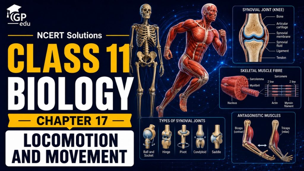
The chapter Locomotion and Movement introduces students to the different types of movements found in living organisms and explains how the human body produces movement through the coordinated action of muscles, bones, and joints. It covers amoeboid movement, ciliary movement, muscular movement, the human skeletal system, types of joints, skeletal muscles, and the mechanism of muscle contraction.
Students learn about different types of movements, including amoeboid, ciliary, and muscular movements, along with their examples and mechanisms. The chapter also explains the human skeletal system, including the axial and appendicular skeleton, major bones, skeletal structure, and the role of bones in providing support, protection, and movement.
Students are introduced to the detailed structure of skeletal muscles, including muscle fibres, myofibrils, sarcomeres, actin, myosin, Z-line, A-band, I-band, H-zone, and M-line. The chapter explains the mechanism of muscle contraction through the sliding filament theory and describes the important roles of calcium ions, troponin, tropomyosin, $\text{ATP}$, actin, and myosin.
Class 11 Biology Chapter 17 Overview
Students learn about the human skeletal system, including the skull, vertebral column, thoracic cage, pectoral girdle, pelvic girdle, and bones of the limbs. The chapter also explains different types of joints, including fibrous, cartilaginous, and synovial joints, along with examples such as shoulder, hip, knee, and elbow joints.
The chapter also covers the structure and function of skeletal muscles and explains how muscle fibres contract and relax. Students study the arrangement of actin and myosin filaments within sarcomeres and understand how their interaction produces muscular movement. The sliding filament mechanism is particularly important for understanding how skeletal muscles generate force and movement.
The chapter further discusses important muscular and skeletal disorders, including myasthenia gravis, muscular dystrophy, tetany, arthritis, osteoporosis, and gout. A clear understanding of Locomotion and Movement helps students develop a strong foundation in human physiology, skeletal structure, muscular contraction, biomechanics, and body movement, making it an important chapter for NEET preparation.
NCERT Solutions for Class 11 Biology
Chapter 17 – Locomotion and Movement
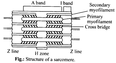
Question 17.1. Draw the diagram of a sarcomere of skeletal muscle showing different regions.
Solution:
Question 17.2. Define sliding filament theory of muscle contraction.
Solution:
According to sliding filament theory of muscle contraction, the actin and myosin filaments slide past each other with the help of cross-bridges to reduce the length of the sarcomeres.
Question 17.3. Describe the important steps in muscle contraction.
Solution:
Mechanism of muscle contraction is explained by sliding filament theory which states that contraction of a muscle fibre takes place by the sliding of the thin filaments over the thick filaments. As a nerve impulse reaches the terminal end of the axon, synaptic vesicles fuse with the axon membrane and release a chemical transmitter, acetylcholine and binds to receptor sites of the motor end plate. When depolarisation of the motor end plate reaches a certain level, it creates an action potential. An action potential (impulse) passes from the motor end plate over the sarcolemma and then into the T-tubules and sarcoplasmic reticulum and stimulates the sarcoplasmic reticulum to release calcium ions into the sarcoplasm. The calcium ions bind to troponin causing a change in its shape and position. This in turn alters shape and the position of tropomyosin, to which troponin binds. This shift exposes the active sites on the F-actin molecules. Myosin cross-bridges are then able to bind to these active sites. The heads of myosin molecules project laterally from thick myofilaments towards the surrounding thin myofilaments. These heads are called cross bridges. The head of each myosin molecule contains an enzyme myosin ATPase. In the presence of myosin ATPase, $\text{Ca}^{2+}$ and $\text{Mg}^{2+}$ ions, $\text{ATP}$ breaks down into $\text{ADP}$ and inorganic phosphate, releasing energy in the head.
Energy from $\text{ATP}$ causes energised myosin cross bridges to bind to actin.

The energised cross-bridges move, causing thin myofilaments to slide along the thick myofilaments.
Question 17.4. Write true or false. If false change the statement so that it is true.
(a) Actin is present in thin filament.
(b) H-zone of striated muscle fibre represents both thick and thin filaments.
(c) Human skeleton has 206 bones.
(d) There are 11 pairs of ribs in man.
(e) Sternum is present on the ventral side of the body.
Solution:
(a) True
(b) False – H-Zone of striated muscle fibres represents only thick filaments.
(c) True
(d) False – There are 12 pairs of ribs in man.
(e) True
Question 17.5. Write the differences between:
(a) Actin and Myosin
(b) Red and White muscles
(c) Pectoral and Pelvic girdle
Solution:
(a) Actin filaments and myosin filaments can be differentiated as follows:
| Actin filaments (Thin) | Myosin filaments (Thick) |
|---|---|
| Composed of F-actin, troponin, and tropomyosin. | Composed of heavy meromyosin (HMM) and light meromyosin (LMM). |
| Found in I-band and extend into A-band. | Restricted to A-band (H-zone). |
(b) Differences between red muscle fibres and white muscle fibres:
| Red muscle fibres | White muscle fibres |
|---|---|
| Contain high amount of myoglobin (red coloured pigment). | Contain low amount of myoglobin. |
| High number of mitochondria, aerobic respiration. | Fewer mitochondria, anaerobic respiration. |
| Do not fatigue easily. | Fatigue faster. |
(c) Differences between pectoral and pelvic girdles:
| Pectoral girdle | Pelvic girdle |
|---|---|
| Connects upper limbs to the axial skeleton (consists of clavicle and scapula). | Connects lower limbs to the axial skeleton (consists of coxal bones). |
| Provides high mobility to arms. | Provides strong support and weight-bearing to legs. |
Question 17.6. Match Column I with Column II:
| Column I | Column II |
|---|---|
| (a) Smooth muscle | (i) Myoglobin |
| (b) Tropomyosin | (ii) Thin filament |
| (c) Red muscle | (iii) Sutures |
| (d) Skull | (iv) Involuntary |
Solution:
(a) – (iv), (b) – (ii), (c) – (i), (d) – (iii)
Question 17.7. What are the different types of movements exhibited by the cells of human body?
Solution:
The cells of human body show three types of movements: amoeboid, ciliary and muscular.
Amoeboid movements: These are found in leucocytes of blood and phagocytes of certain body organs. In such cells, movements are brought with the help of temporary finger-like cytoplasmic projections, called pseudopodia or false feet. So it is also called pseudopodial movement. These pseudopodia are formed by flow of cytoplasm, called cyclosis, and cytoskeletal structures like microfilaments.
Ciliary movements: Large number of our internal tubular organs are lined by ciliated epithelium. For instance, the cilia of the cells lining the trachea, oviducts and vasa efferentia propel dust particles, eggs and sperms respectively by their coordinated movements in specific directions in these organs.
Muscular movements: These are brought about by the action of skeleton, joints and muscles. These are of two types: movements of body parts and locomotion.
Question 17.8. How do you distinguish between a skeletal muscle and a cardiac muscle?
Solution:
| Skeletal muscle | Cardiac muscle |
|---|---|
| Voluntary in function. | Involuntary in function. |
| Unbranched, cylindrical fibers with multiple nuclei. | Branched fibers with intercalated discs and single/binucleated cells. |
| Attached to bones. | Found exclusively in the heart walls. |
Question 17.9. Name the type of joint between the following:
(a) atlas/axis
(b) carpal/metacarpal of thumb
(c) between phalanges
(d) femur/acetabulum
(e) between cranial bones
(f) between pubic bones in the pelvic girdle
Solution:
(a) Pivot joint
(b) Saddle joint
(c) Hinge joint
(d) Ball and socket joint
(e) Fibrous joint (sutures)
(f) Cartilaginous joint (pubic symphysis)
Question 17.10. Fill in the blank spaces:
(a) All mammals (except a few) have ……. cervical vertebra.
(b) The number of phalanges in each limb of human is …….
(c) Thin filament of myofibril contains two ‘F’ actins and two other proteins namely ……. and …….
(d) In a muscle fibre $\text{Ca}^{2+}$ is stored in …….
(e) ……. and ……. pairs of ribs are called floating ribs.
(f) The human cranium is made of ……. bones.
Solution:
(a) 7
(b) 14
(c) tropomyosin, troponin
(d) sarcoplasmic reticulum
(e) $11^{\text{th}}$ and $12^{\text{th}}$
(f) 8
Why Class 11 Biology Chapter 17 Matters in NEET
Class 11 Biology Chapter 17: Locomotion and Movement is highly important for NEET because it explains the mechanisms by which organisms and body parts move. The chapter covers important concepts such as types of movement, skeletal muscles, the human skeletal system, joints, muscle contraction, sliding filament theory, and disorders related to the muscular and skeletal systems. NEET frequently includes NCERT-based questions on the structure and functions of muscles, bones, joints, sarcomeres, actin and myosin, muscle contraction, and the role of calcium ions and $\text{ATP}$. A thorough understanding of this chapter helps students build a strong foundation in human physiology, biomechanics, muscular coordination, and the movement of the human body while improving their performance in the examination.
Preparation Tips for Class 11 Biology Chapter 17
Begin by understanding the different types of movement, including amoeboid movement, ciliary movement, and muscular movement. Learn suitable examples and understand how these movements occur in different organisms and body parts.
Study the human skeletal system carefully, including the axial and appendicular skeleton. Pay special attention to the major bones of the skull, vertebral column, thoracic cage, pectoral girdle, pelvic girdle, and limbs. Practise identifying important bones from NCERT diagrams.
Understand the structure of skeletal muscle fibres in detail. Focus on myofibrils, sarcomeres, actin, myosin, Z-lines, A-band, I-band, H-zone, and the M-line. Learn the arrangement of thick and thin filaments and how they change during muscle contraction.
Study the mechanism of muscle contraction thoroughly, especially the sliding filament theory. Understand the roles of calcium ions, troponin, tropomyosin, $\text{ATP}$, actin, and myosin. Pay attention to the sequence of events involved in cross-bridge formation and relaxation of the muscle.
FAQs
1. What are the most important topics in Class 11 Biology Chapter 17?
The most important topics include types of movement, human skeletal system, bones and joints, skeletal muscles, muscle fibre structure, sarcomere, actin and myosin, sliding filament theory, muscle contraction, and disorders of the muscular and skeletal systems. These topics are frequently tested in NEET.
2. What is locomotion?
Locomotion is the movement of an organism from one place to another. It is generally associated with movement of the entire organism, whereas movement may involve only a part of the body.
3. What are the major types of movement?
The major types of movement discussed in this chapter are amoeboid movement, ciliary movement, and muscular movement. Different organisms and cells use different mechanisms to produce movement.
4. What is a sarcomere?
A sarcomere is the functional unit of a myofibril in a skeletal muscle. It extends between two successive Z-lines and contains the arrangement of actin and myosin filaments responsible for muscle contraction.
5. What is the sliding filament theory?
The sliding filament theory explains muscle contraction by describing how actin filaments slide over myosin filaments, causing the sarcomere to shorten. The filaments themselves do not significantly shorten; instead, their relative positions change during contraction.
6. What is the role of calcium ions in muscle contraction?
Calcium ions play an essential role in initiating muscle contraction. They bind to troponin, causing a change in the position of the troponin-tropomyosin complex and exposing the active sites on actin for interaction with myosin.
7. What are the different types of joints?
Joints are broadly classified as fibrous, cartilaginous, and synovial joints. Synovial joints include ball-and-socket, hinge, pivot, gliding, and saddle joints, each allowing different types and ranges of movement.
8. What are some important disorders mentioned in this chapter?
Important disorders include myasthenia gravis, muscular dystrophy, tetany, arthritis, osteoporosis, and gout. Students should learn their basic causes and characteristics according to NCERT.